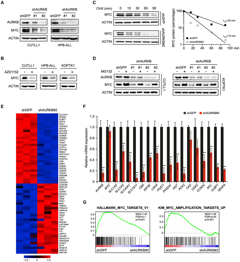
Figure 2. AURKB Inhibition Attenuates the MYC Protein Accumulation and Transcriptional Activity.
(A) AURKB was depleted by specific shRNAs in T-ALL cells as indicated. AURKB and MYC proteins were analyzed by immunoblot, with ACTIN as a loading control. shGFP, control shRNA targeting GFP.
(B) Immunoblots of MYC and ACTIN in T-ALL cells treated with 10 nM AZD1152 for 24 h.
(C) Time-course analysis of MYC protein levels in AURKB-depleted CUTLL1 cells. MYC proteins were quantified and plotted on the right.
(D) AURKB-depleted CUTLL1 and KOPTK1 cells were treated with MG132 (10 μM) for 6 h before harvest. AURKB and MYC were analyzed by immunoblot, with ACTIN as a loading control.
(E) A heatmap depicting top 30 down- and upregulated genes upon AURKB depletion in CUTLL1 cells (Q < 0.05).
(F) qRT-PCR analysis of representative MYC-induced target genes in CUTLL1 cells upon AURKB depletion. Data shown represent the means (±SD) of biological triplicates. *p < 0.05, **p < 0.01, unpaired two-tailed Student’s t test.
(G) Gene set enrichment analysis of MYC target gene sets in the expression profiles of CUTLL1 cells expressing AURKB shRNA (http://software.broadinstitute.org/gsea).
See also Figure S1.

