
Figure 2.
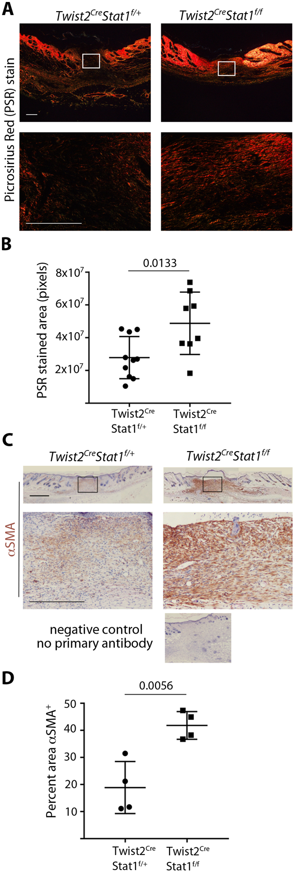
Mesenchymal Stat1 deletion leads to more collagen deposition and increased αSMA expression during wound healing. (A) Collagen deposition in the wound center at day 7 of healing, stained with picrosirius red (PSR) and visualized under polarized light. Boxed area in upper panels is magnified in lower panels. (Scale bar = 500μm). (B) Quantification of PSR stained area in the wound center as shown in A, with n = 8–10 wounds per genotype. (C) αSMA protein expression in the wound center at day 7 of healing, stained with αSMA immunohistochemistry (brown) and hematoxylin counterstain. Boxed area in upper panels is magnified in lower panels. (Scale bar = 1mm) (D) Quantification of αSMA staining as a percentage of the wound bed area, with n = 4 wounds per genotype. Student’s t-test was used for statistical analysis.