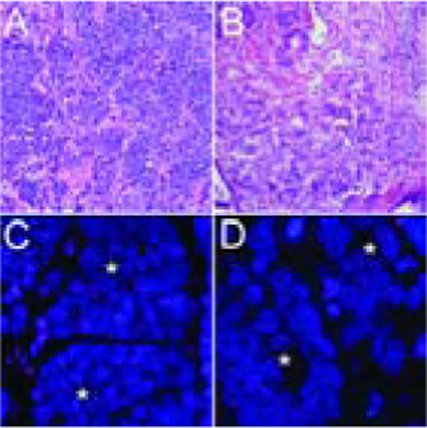
Figure 2. Telomere-specific FISH in a case with small cell carcinoma and adenocarcinoma components.

In a representative case, the telomere signals are robust in the cancer cells (*) from both the (A, C) small cell carcinoma and the (B, D) concurrent adenocarcinoma. In all images, the DNA is stained with DAPI (blue) and telomere DNA is stained with the Cy3-labeled telomere-specific PNA probe (red). The centromere DNA, stained with the FITC-labeled centromere-specific peptide nucleic acid probe, has been omitted from the image to emphasize the differences in telomere lengths. Original magnification × 400.
