
Figure 3. Telomere length status by histologic diagnosis.
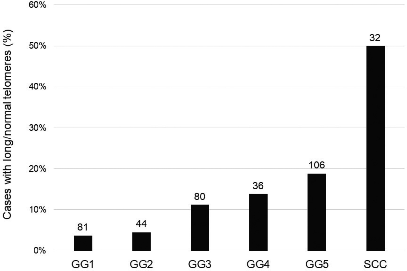
There is a step-wise increase in the prevalence of normal/long telomere lengths among adenocarcinoma cases with increasing Grade Group category. Prostatic small cell carcinoma cases show the highest prevalence of normal/long telomere lengths. The numbers over each bar represent the total number of cases examined for each category.