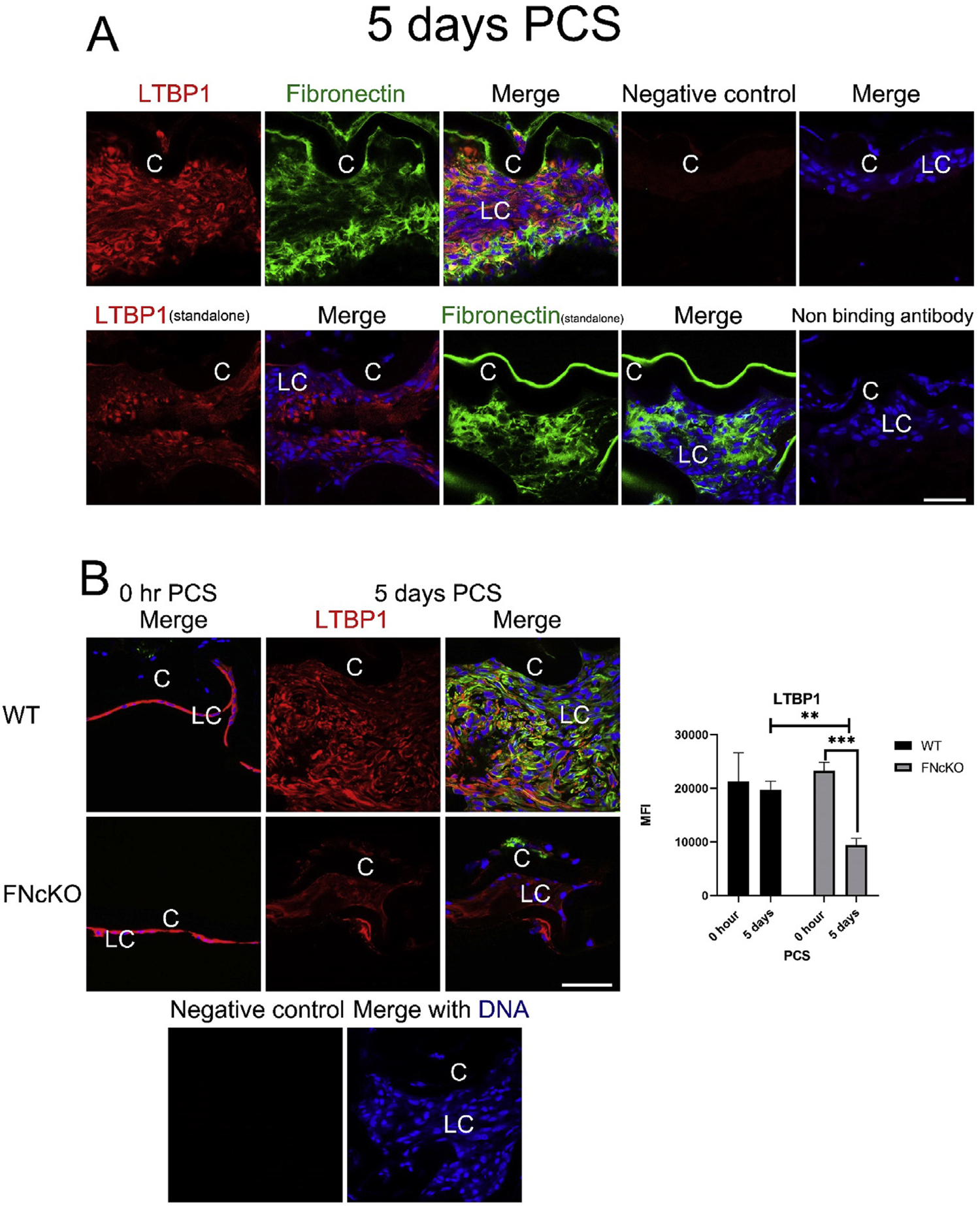
Fig. 6. LCs are associated with latent TGFβ binding protein at 5 days PCS, and this is highly attenuated in FNcKO LCs.

(A) At 5 days PCS, WT LCs are associated with robust levels of cell-associated fibronectin and LTBP1. Fibronectin (green) and LTBP1 (red) merged with DNA detected by Draq5 (blue). (B) At 0 h PCS, appreciable levels of LTBP1 protein are detected in both WT and FNcKO LCs whereas αSMA protein levels are low. However, by 5 days PCS, WT LCs maintain the robust levels of ECM-associated LTBP1 whereas extracellular deposition of LTBP1 around FNcKO LCs is greatly attenuated (**P = 0.006) compared to WT and is even reduced compared to 0 h PCS (***P ≤ 0.001). αSMA (green) and LTBP1 (red) merged with DNA detected by Draq5 (blue). Scale bars: 35 μm. LC, remnant lens epithelial cells/lens cells; C, lens capsule. All experiments had N = 3. Values are expressed as mean ± SEM. Asterisks (*) indicate statistically significant difference in MFI between WT and FNcKO at a time PCS or between two PCS time points.
