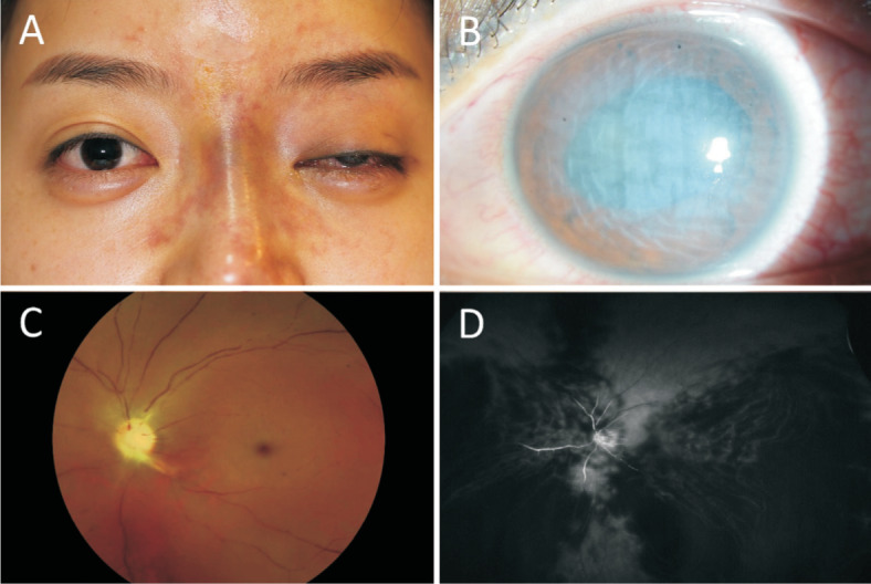
Figure 1. Clinical findings at initial presentation.

A: Ptosis with incomplete ophthalmoplegia in the left eye and nasal skin discoloration; B: Corneal edema with anterior chamber inflammation on slit lamp biomicroscopy; C: Multiple emboli in arterial vascular arcade and whitened retina with cherry red spot on fundus photography; D: FA showing severely diminished retinal and choroidal perfusion with diffuse non-perfused area, even in the late phase.
