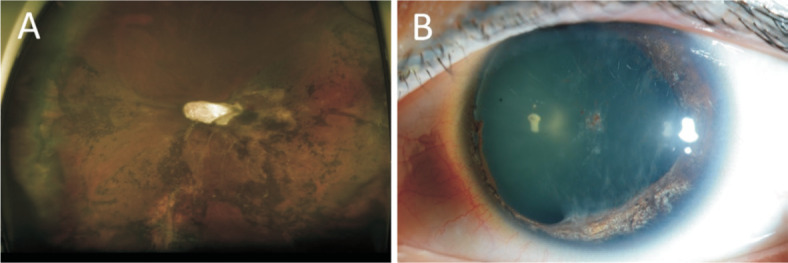
Figure 3. Fundus and anterior segment findings 3y post treatment.

A: Diffuse retinal atrophy with thick tractional membrane are observed on fundus photography; B: Iris atrophy and cataract, which resulted from anterior segment ischemia, were aggravated.
