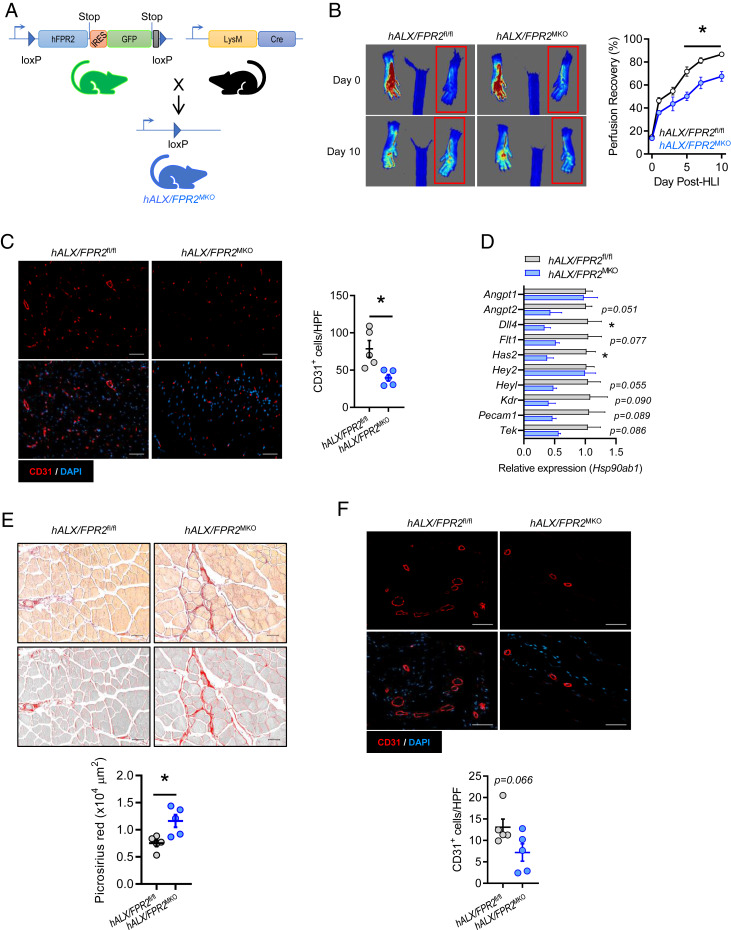
Fig. 4.
Myeloid ALX/FPR2 has a cell-autonomous role in ischemic revascularization. (A) Schematic representation of the breeding strategy to generate myeloid-specific ALX/FPR2 KO (hALX/FPR2MKO) mice. The murine Fpr2 gene was replaced with human FPR2 (hFPR2) with a GFP reporter preceded by an internal ribosome entry site (IRES) to generate GFP+ FPR2 floxed mice (hFPR2fl/fl). These mice were then bred with LysM-cre mice to produce mice with myeloid-specific deletion of FPR2 (hALX/FPR2MKO). (B) Representative images (Left) and quantification (Right) of laser speckle contrast perfusion imaging in hALX/FPR2fl/fl and hALX/FPR2MKO mice following HLI. The red box denotes the ischemic region of interest. n = 8 to 10 per group. *P < 0.05 at the indicated time points, two-way ANOVA with Sidak multiple comparison test of hALX/FPR2MKO vs. hALX/FPR2fl/fl. (C) Representative immunofluorescence images (Left) and quantification (Right) of CD31+ cells in ischemic gastrocnemius muscle of hALX/FPR2fl/fl and hALX/FPR2MKO mice on day 14 of HLI. n = 5 per group. (D) Expression of vascular-related genes (relative to Hsp90ab1) in ischemic gastrocnemius muscle of hALX/FPR2fl/fl and hALX/FPR2MKO mice on day 3 of HLI. n = 3 per group. (E) Representative brightfield and grayscale images (Top) and quantification (Bottom) of picrosirius red-stained ischemic hamstrings of WT and Alx/Fpr2MKO mice on day 14 of HLI. n = 5 per group. (F) Representative immunofluorescence images (Top) and quantification (Bottom) of CD31+ cells in cutaneous wounds of hALX/FPR2fl/fl and hALX/FPR2MKO mice on day 3 after wounding. n = 5 per group. Data are presented as mean ± SEM. In C–F, *P < 0.05, unpaired Student’s t test. (Scale bars: 50 µm.)

