Fig. 1.
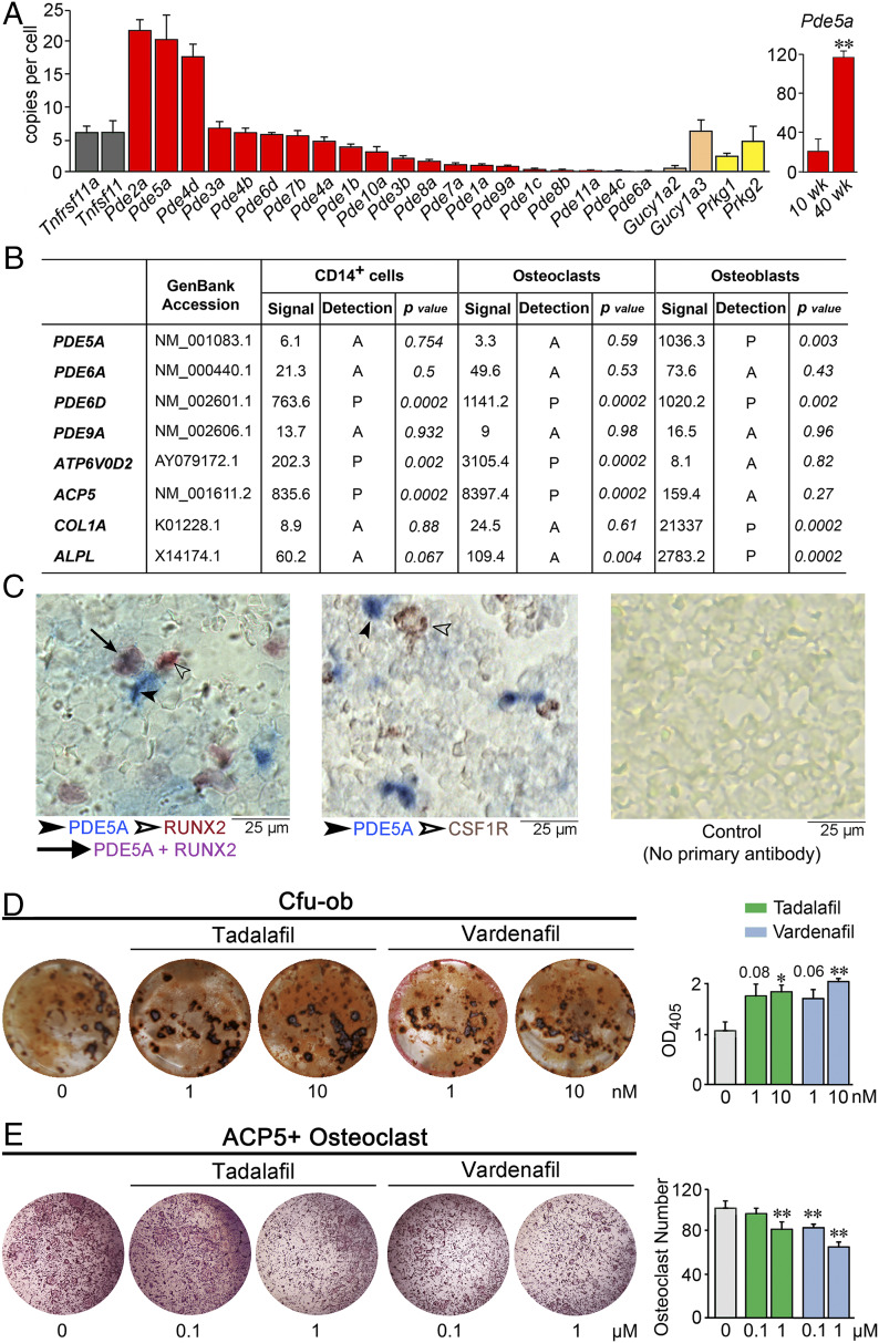
Expression and in vitro actions of PDE5A inhibitors tadalafil and vardenafil. (A) TaqMan-based PCR using bone RNA showing the expression of 20 murine PDE isoforms, soluble guanylate cyclase isoforms (Gucy1a2 and Gucy1a3), protein kinase G isoforms (Prkg1 and Prkg2), as well as Tnfrsf11a and Tnfsf11 (controls). SYBR Green-based PCR using bone RNA from 10- and 40-wk-old mice showing the expression of Pde5a. Data are expressed as copy number per cell (normalized to Tubulin, Rps11, Actb, and/or Gapdh). Data are mean ± SEM. n = 3 biological replicates for TaqMan; n = 4 biological replicates for SYBR Green. (B) Data obtained from Human Genome U133 2.0 GeneChip Arrays (Affymetrix). The presence of transcripts was determined from the signal of perfect matched and mismatched probe pairs in each probe set, with statistical confidence (P value) indicated. Characteristic highly expressed osteoclastic and osteoblastic transcripts are also included as controls. (C) Photomicrographs showing immune labeling of PDE5A (blue, solid arrowhead) and RUNX2 (Left) or CSF1R (Middle) (brown, hollow arrowhead) in mouse bone marrow (colocalization is shown in purple, solid arrow), together with a control image without primary antibody (Right) (details in Methods). (D) Effect of tadalafil and vardenafil on Cfu-ob in 21-d cultures of bone marrow stromal cells isolated from marrow of 10-mo-old male mice in differentiating medium, shown as representative alizarin red-positive Cfu-ob and mean ± SEM absorbance of extracted dye per well (in triplicate). (E) Effect of tadalafil and vardenafil on tartrate-resistant acid phosphatase-positive (ACP5+) osteoclasts at 5 d following the incubation of bone marrow hematopoietic stem cells with RANKL and M-CSF, shown as representative plates and mean ± SEM ACP5+ osteoclast number per well (in triplicate). Statistics for D and E: unpaired two-tailed Student’s t test; *P < 0.05, **P < 0.01, or as shown.

