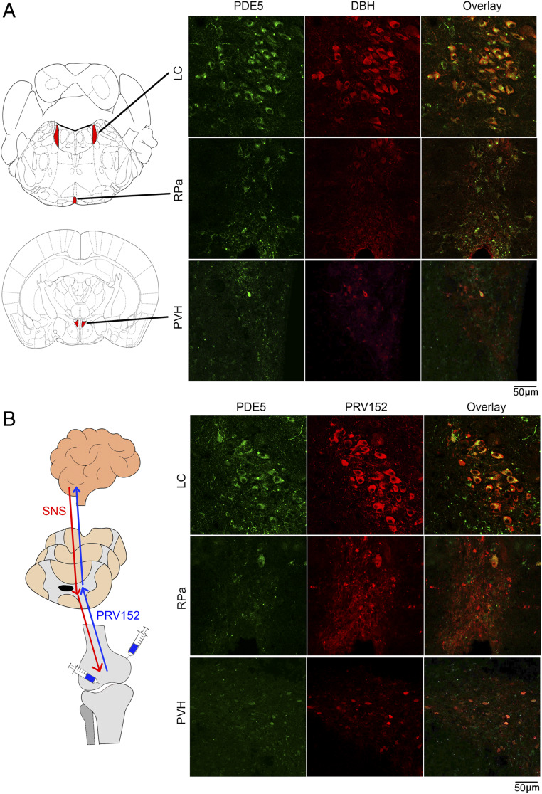
Fig. 2.
Localization of PDE5A in sympathetic neurons in three brain regions. (A) Representative confocal photomicrographs showing the colocalization (yellow) of PDE5A (green) and DBH (red) immunolabeling in a number of neurons of the locus coeruleus (LC), raphe pallidus (RPa), and paraventricular nucleus of the hypothalamus (PVH). Also shown is the map of brain areas. (B) Retrograde transsynaptic virus-mediated tract tracing using a pseudorabies virus strain, PRV152, that expresses EGFP under control of the human cytomegalovirus immediate-early promoter. PRV152 was injected into the metaphysis or subperiosteally (shown as schematic) in live anesthetized mice at 6 d before sacrifice. Brain regions were dissected and processed for PDE5A (green) and EGFP (red) immunohistochemistry. The virus traversed from bone via the sympathetic nervous system to the three brain regions, LC, Rpa, and PVH, where it colocalized with PDE5A (yellow). Refer to SI Appendix, Fig. S1.

