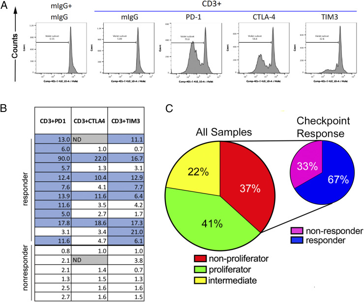
Fig. 4.
Impaired T cell proliferation in response to TCR ligation can be reversed by immune checkpoint blockade in a subset of patients. (A) A representative “checkpoint responder” showing CTV dilution with the addition of antibodies against PD-1, CTLA-4, and TIM3. (B) A summary of the responses of all nonproliferators to checkpoint molecule blockade. Numbers within boxes represent the fold-change in proliferation, as determined by the proliferation measured with anti-CD3 and PD-1, CTLA-4, or TIM3 antibody treatment divided by the proliferation with anti-CD3 + mIgG. Blue-shaded boxes indicate checkpoint responders, defined as samples with at least a five-fold increase in proliferation value when comparing anti-CD3 + checkpoint antibody to anti-CD3 + mIgG. (C) Summary of results from the the patient cohort assayed for T cell function (n = 49).

