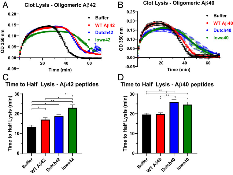
Fig. 3.
HCAA-type Aβs increase resistance to fibrinolysis compared to WT Aβ. (A and B) Fibrin clot formation and degradation were assessed by measuring time-dependent turbidity changes at 350 nm. These experiments involved incubation of fibrinogen, tPA, plasminogen, and the absence (black, buffer control only) or presence of WT Aβ (red) or HCAA Aβ peptides (blue for Dutch, green for Iowa). Reactions were initiated by adding thrombin. Time-dependent turbidity plots show the effect of WT Aβ42 and HCAA Aβ42 (A) and WT Aβ40 and HCAA Aβ40 (B) on thrombosis and fibrinolysis. (C) Time to half-lysis times for WT Aβ42, Dutch42, and Iowa42 are prolonged compared to control. Clot lysis time of Iowa42 was significantly more delayed than WT Aβ42. (**P < 0.01; *P < 0.05; n = 3). (D) While WT Aβ40 did not have any effect on fibrin clot lysis, Dutch40 and Iowa40 significantly delayed fibrinolysis compared to buffer alone and WT Aβ40. (**P < 0.01; *P < 0.05; n = 3). Statistical analyses were performed using one-way ANOVA followed by Tukey’s post hoc test. Bar graphs represent mean ± SEM of ≥3 separate experiments.

