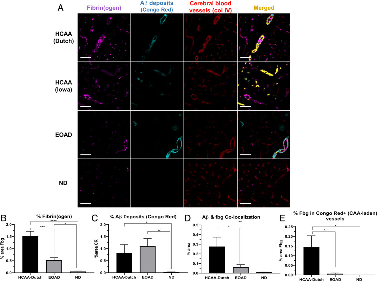
Fig. 4.
Increased fibrin(ogen) deposits and Aβ-fibrino(gen) codeposition in HCAA patients’ occipital cortex. Representative 20× images of brain sections were probed with antibodies against fibrin(ogen) (fbg, magenta), congophillic Aβ deposits using Congo Red dye (CR) (cyan), and blood vessels (col IV, red). (A) Occipital cortical sections in HCAA-Dutch (n = 5) and -Iowa (n = 1) type patient brains show abundant intra- and extravascular fibrin(ogen) deposits (magenta) and high amounts of vascular Aβ (cyan) around cerebral blood vessels (red), demarcated by basement membrane collagen IV, which frequently overlapped (Merged, yellow) at sites of CAA. (B and C) Quantification of percent area of target protein reveals elevated levels of fibrin(ogen) and Aβ deposits, respectively. (D and E) Confocal analysis shows elevated colocalization between Aβ and fibrin(ogen) in HCAA-Dutch brains, with particularly higher levels of fibrin(ogen) codeposition in CAA-laden vessels (Congo Red-positive), compared to EOAD (n = 5) and ND (n = 7) brains. Due to the limited amount of HCAA-Iowa (n = 1) individuals available in our study, it was not included in our IF quantification analysis. Statistical analyses were performed using one-way ANOVA followed by Tukey’s post hoc test. ****P < 0.0001; ***P < 0.001; **P < 0.01; *P < 0.05. (Scale bars: 100 µm.)

