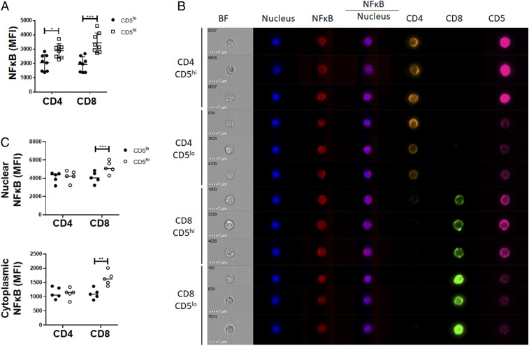
Fig. 5.
The CD5hi and CD5lo fractions of peripheral T cells show differential pool of cytoplasmic NF-κB. (A) gMFI of NF-κB in peripheral CD4 and CD8 T cells isolated from lymph nodes (n = 8 from 3 independent experiments). (B) Purified WT T cells were stained and analyzed by imaging flow cytometry. Screenshot of dashboard from IDEAS analysis from data collected on Amnis ImageStream. (C) Mean NF-κB expressed as a ratio of CD5hi to CD5lo within CD4 and CD8 peripheral resting T cells (n = 5 from 1 independent experiment). *P < 0.05; **P < 0.01; ***P < 0.001; anything not marked is not statistically significant.

