Fig. 6.
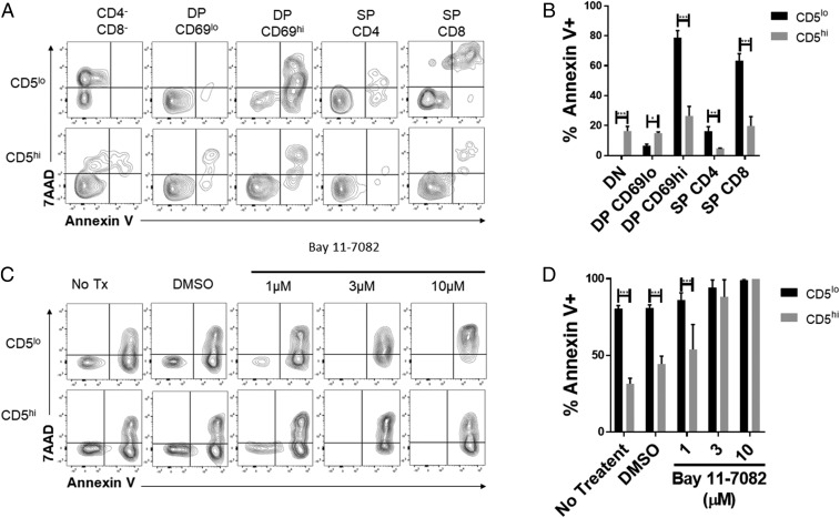
CD5 expression confers thymocyte survival advantage. (A) Thymocytes from B6 mice were cultured ex vivo for 24 h. Cells were stained and gated as in SI Appendix, Fig. S2A, together with Annexin V and 7AAD to assess cell death. The populations were grouped based on top and bottom 20% of CD5 expression within each population. Representative flow plots are shown. (B) The frequencies of Annexin V+ cells within the top and bottom 20% of CD5 expressers are shown (n = 4 from 1 experiment is shown and n = 3 from a second independent experiment). (C) Thymocytes from B6 mice were cultured ex vivo for 15 h with or without varying Bay 11-7082 doses. Cells were stained and gated as in SI Appendix, Fig. S2, together with Annexin V and 7AAD to assess cell death. Representative flow plots are shown for the double-positive CD69hi population. (D) The frequencies of Annexin V+ cells within the top and bottom 20% of CD5 expressers of the double-positive CD69hi population are shown (n = 3 from 1 independent experiment). *P < 0.05; **P < 0.01; ***P < 0.001; anything not marked is not statistically significant.

