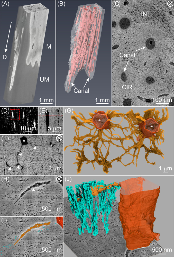
Fig. 1.
The tenocyte network in a highly mineralized zone of TLT from a tendon prepared by chemical fixation and stained with osmium tetroxide alone. (A) Three-dimensional reconstruction of micro-CT data from the more heavily mineralized distal end of the leg tendon. This region contains both mineralized (radiopaque, M) and unmineralized (darker gray, UM) zones. The arrow points toward the distal end (D) of the tendon. (B) The mineralized zone in A contains multiple unmineralized canals (pink) principally oriented in the tendon longitudinal direction. (C) High-resolution micro-CT image of a representative transverse section from a fully mineralized zone of the tendon reveals circumferential regions (CIRs) with qualitatively lower porosity surrounding the canals and interstitial tissue (INT) with higher porosity between CIRs. (D) Confocal image of a longitudinal section in the mineralized zone of a rhodamine-stained tendon showing tenocyte lacunae and canaliculi (white streaks and small white dots/lines, respectively). An area framed in red is enlarged and shown by energy selective backscattered (EsB) imaging in E where lacunae and canaliculi are more apparent. (F) EsB image of a transverse section (marked by the red dashed line in E) showing two tenocyte lacunae (*) and radiating canaliculi (arrowheads) surrounding mineralized collagen fibril bundles. (G) FIB-SEM 3D reconstruction of a volume of F illustrating the two tenocyte lacunae (tangerine) and their associated canaliculi (gold). (H) High-resolution SE image of a tendon section showing a portion of a cell lacuna, canaliculus (arrowhead) and fine pores, highlighted in dark tangerine, gold, and turquoise, respectively in I. (J) Three-dimensional rendering corresponding to H and I of the tenocyte lacuno-canalicular network intersecting with a FIB-SEM background image plane and demonstrating the numerous secondary channels (turquoise) branching from a canaliculus (gold) and disposed primarily in the longitudinal tendon direction. The tenocyte lacuna is defined by a contoured surface (tangerine). The circled cross in C, F, and H denotes the view along the longitudinal direction of the tendon.

