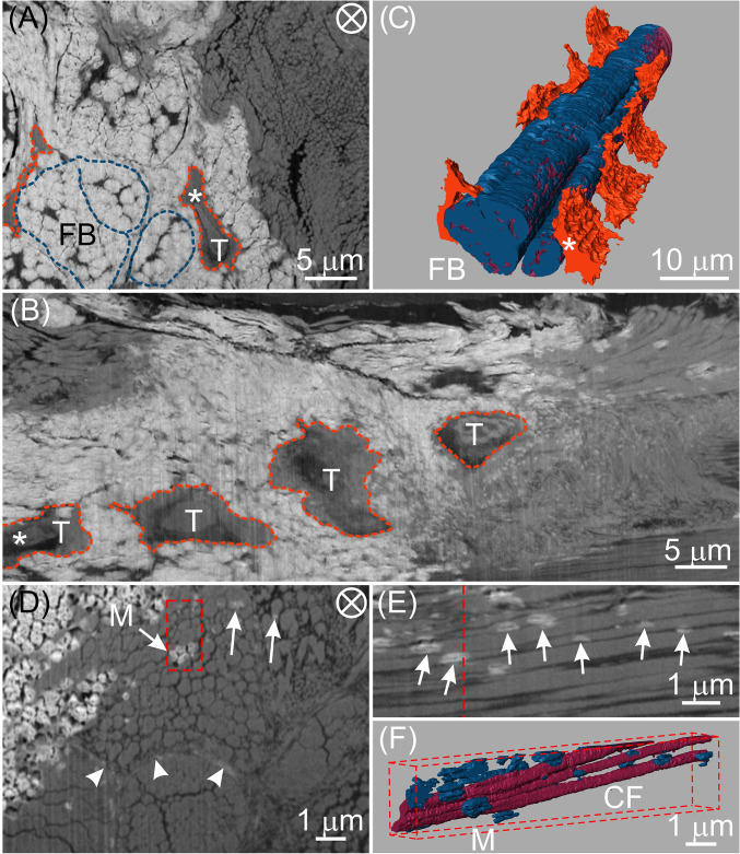
Fig. 2.
FIB-SEM images and corresponding 3D reconstructions of tenocyte lacunae, collagen fibrils, and mineral deposits at an interface between mineralized and unmineralized avian tendon zones. Images are from the same specimen, prepared by cryomicrotomy and HPF. AFS followed these procedures and the sample was stained with uranyl acetate alone. (A) EsB image of a transverse section of a typical tendon volume showing mineralized (white) and unmineralized (gray) zones. Two collagen fibril bundles (FB) and tenocytes (T) may be identified in the mineralized zone and are denoted by dashed blue and tangerine lines, respectively. There are thin, dark structures of unknown nature that appear to separate individual mineralized units within bundles. Such structures may possibly be related to canaliculi surrounding fibril bundles. (B) Longitudinal view obtained by digital processing the full stack of FIB-SEM slices through the same tendon volume represented by the single transverse EsB image in A. Tenocytes (T) within their lacunae are outlined in tangerine. (C) Three-dimensional reconstruction of the FIB-SEM data of the volume of A and B showing tenocyte lacunae (tangerine) adjacent to and aligned along the longitudinal direction of mineralized collagen fibril bundles. Bundles may be distinguished by their component collagen fibrils (wine) and mineral (blue). The same tenocyte (T) in its lacuna has been followed and identified (*) in each of the images, A and B, and their reconstruction, C. (D) High-resolution EsB image showing mineral (arrows, M) within collagen fibril bundles and sheath regions (arrowheads). (E) Longitudinal view obtained by digital processing the full stack of FIB-SEM slices through the same tendon volume represented by the transverse EsB image in the red-framed area of D. The red dashed line in E represents the location of the framed area of D. Numerous mineral deposits (arrows) are associated with collagen fibrils and appear predominantly as prolate ellipsoids in shape and elongated in the tendon longitudinal direction. (F) Three-dimensional reconstruction of the volume marked by the red-framed area in D showing several collagen fibrils (CF, wine) and their associated discrete mineral deposits (M, blue) that vary in length and thickness. As in E, the longitudinal view reveals numerous mineral deposits interrelated with several collagen fibrils, many of which are separated by dark, narrow spaces. The circled cross in A and D denotes the view along the longitudinal direction of the tendon.

