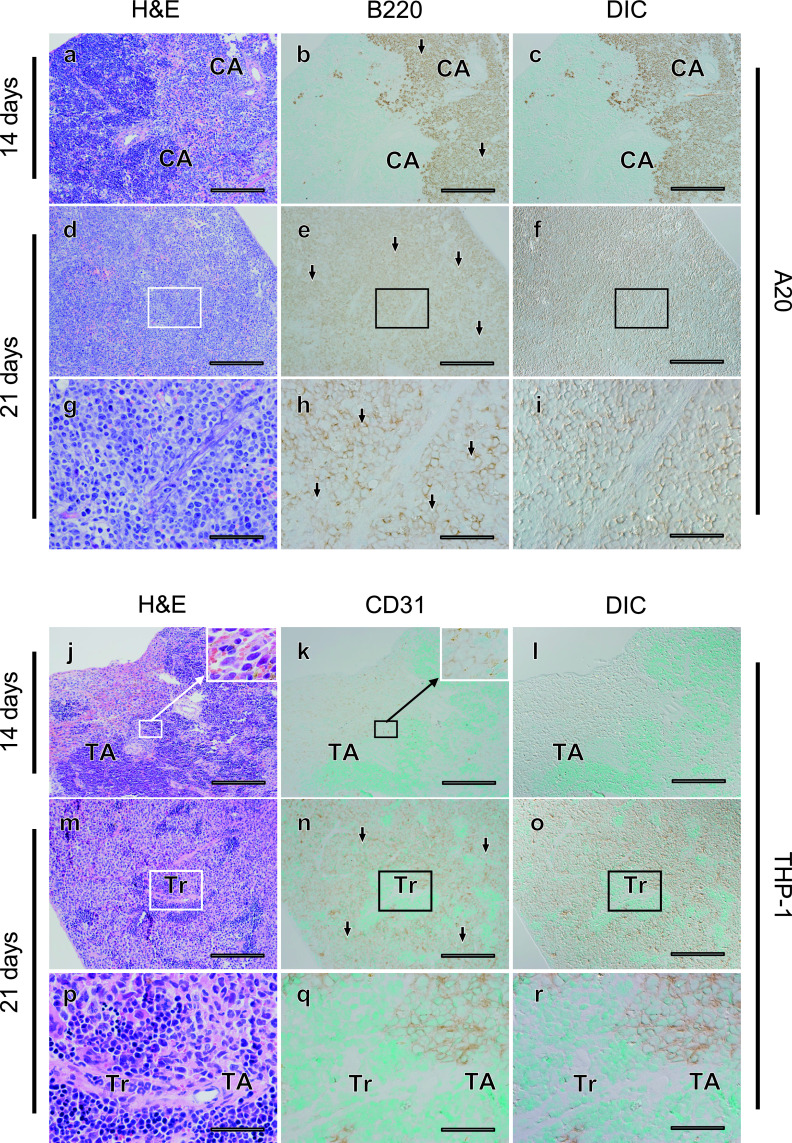
Fig. 2.
Massive infiltration of tumor cells into the spleen parenchyma was commonly observed in the A20 and THP-1 leukemia mouse models, but infiltration into connective tissues was observed only in the A20 model. Fourteen (a–c, j–l) or 21 (d–f, m–o) days after transplantation, histological changes in spleen tissues were evaluated in serial sections (a–c, d–f, j–l, m–o) using hematoxylin-eosin (H&E) staining (a, d, j, m) and tumor cells (arrows) were detected with immunostaining for B220 in A20 leukemia mouse models (b, c, e, f), and for CD31 in THP-1 mouse models (k, l, n, o). Differential interference contrast (DIC) images (c, f, l, o) were acquired in areas where immunostaining for tumor cells was observed (b, e, k, n). Areas indicated with rectangles (d–f, m–o) are magnified (g–i, p–r). CA; central artery, Tr; trabecula, TA; trabecular artery. Bars = 500 μm (a–f, j–o) and 100 μm (g–i, p–r).

