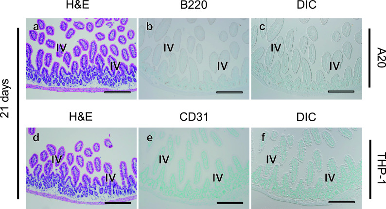
Fig. 6.
No infiltration of A20 or THP-1 cells in the intestine. Twenty one days after transplantation, histological changes in intestinal tissues were evaluated in serial sections (a–c, d–f) using H&E staining (a, d) and tumor cells (arrows) were detected with immunostaining for B220 in A20 leukemia mouse models (b, c), and for CD31 in THP-1 mouse models (e, f). There is no evidence of leukemia invasion into the intestine. IV; intestinal villi. Bars = 500 μm.

