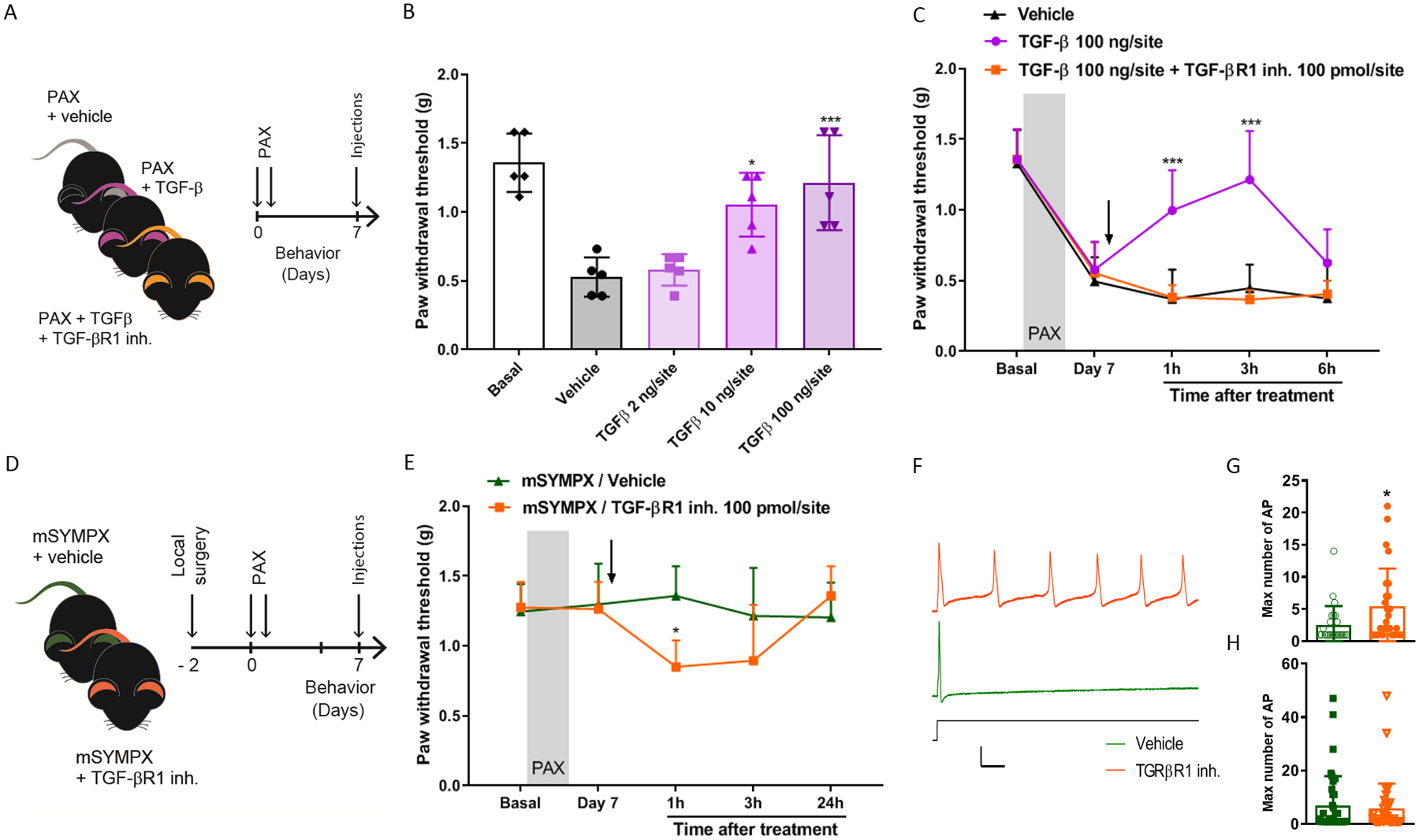
Figure 6. Local sympathectomy accelerates the resolution of paclitaxel-induced mechanical allodynia through anti-inflammatory TGF-β signaling.

(A) Schematic illustration of the experiment showing the timeline of the injections of paclitaxel (PAX), intrathecal injections of recombinant TGF-β protein or TGF-β + TGF-β receptor 1 inhibitor (TGF-βR1 inh.), and the behavioral assay that was carried out at 7 day after paclitaxel. (B) Reversal of paclitaxel-induced mechanical allodynia tested in ipsilateral hind paws in male mice after 3 h of treatment with different doses of recombinant TGF-β (n=5 mice/group, one-way ANOVA showed significant difference between TGF-β and vehicle groups, Group: F4,20 = 14.02, p < 0.001; Turkey post hoc analysis revealed a significant difference at concentrations of 10 ng and 100 ng. *p < 0.05, *p < 0.001). (C) Time course of paclitaxel-induced mechanical allodynia showed that the anti-allodynic effect of TGF-β was abolished by the intrathecal co-injection of the TGF-βR1 inhibitor (n=5 male mice/group, two-way ANOVA showed significant difference between TGF-β and vehicle groups, Group × Time interaction: F8,48 = 5.63, p < 0.001; Bonferroni post hoc analysis revealed a significant difference between groups at 1 and 3 hours. *p < 0.001). (D) Schematic illustration of the experiment showing the timeline of local microsympathectomy (mSYMPX), injections of paclitaxel (PAX), injections of TGF-βR1 inh., and the behavioral assay that was carried out at 7 day after paclitaxel. (E) TGF-βR1 inhibitor also reverses the anti-allodynic effect of mSYMPX on paclitaxel-induced mechanical allodynia in mice (n=5 male mice/group, two-way ANOVA showed significant difference between mSYMPX -SB431542 and -vehicle groups, Group × Time interaction: F4,32 = 3.25, p = 0.024; Bonferroni post hoc analysis revealed a significant difference between groups at 1 hour. *p < 0.05). (F) Sample traces of action potential firing in response to suprathreshold currents in small cells isolated after chemotherapy and mSYMPX, with TGFβ1R inhibitor (orange) or vehicle (green) present during the recording session. Shown is response to stimulus of 2.4 nA which evoked maximum number of action potentials in the TGFβ1R inhibitor cell; and response to stimulus of 3.4 nA in the vehicle treated cell, where it was not possible to evoke more than one action potential. Scale bars 25 msec, 50 mV. (G, H) Quantification of the TGF-βR1 inhibitor effect on the maximum number of action potentials evoked by suprathreshold currents in small-size DRG neurons (G) and large-size DRG neurons (H) isolated from mSYMPX mice treated with paclitaxel (n=28–29 small neurons/group, or 41–43 large neurons per group; Mann-Whitney test, *p < 0.05 compared to vehicle). Additional electrophysiological details can be found in suppl. table 2.
