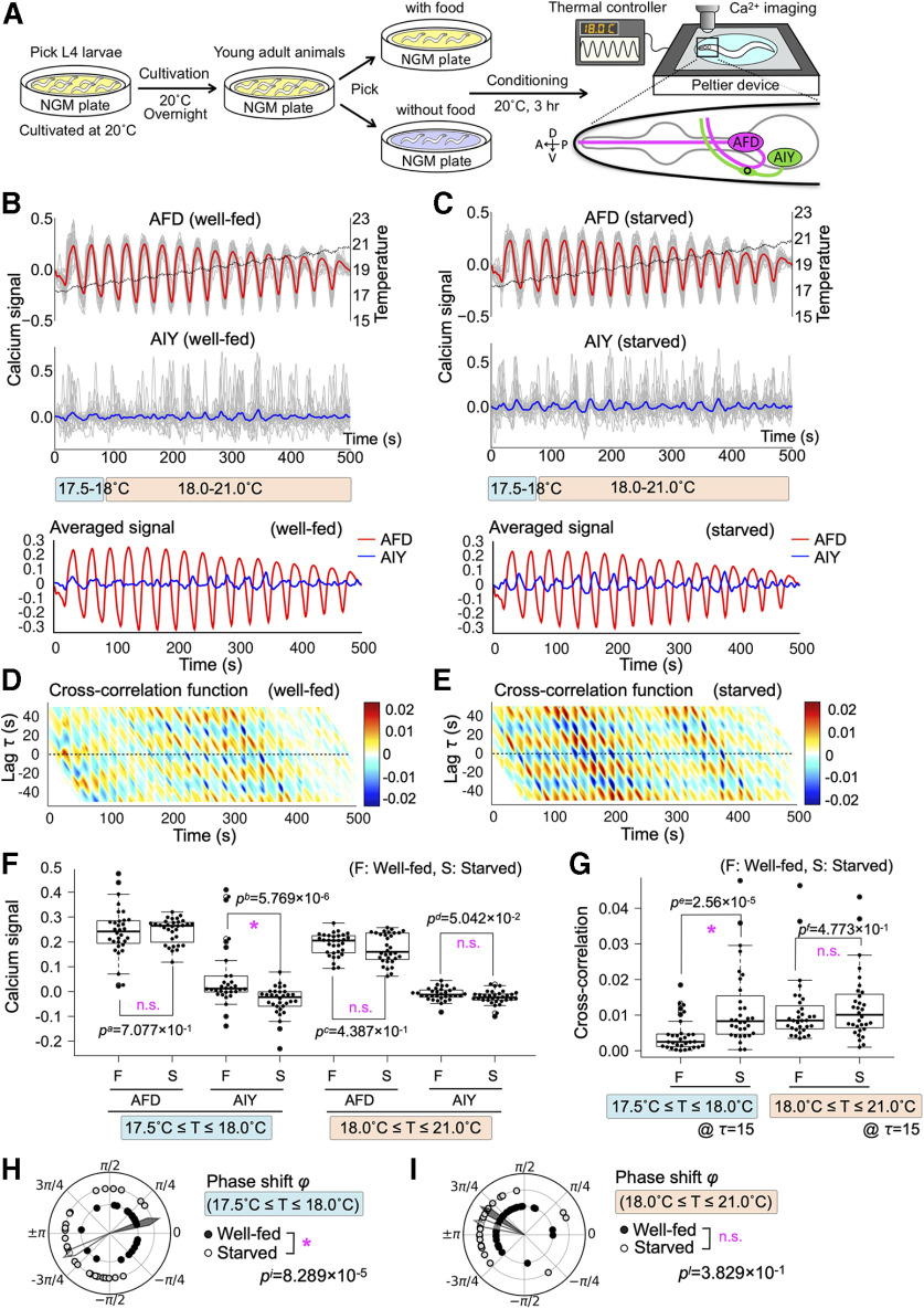
Figure 1.
AFD-AIY temporal interaction in response to oscillatory thermal ramp from 17.5°C to 21.0°C (Osci1721) was altered by starvation. A, The schematics of simultaneous calcium imaging of AFD and AIY under time-varying thermal stimuli. Well-fed and starved animals were conditioned at 20°C for 3 h with and without food, respectively. Genetically encoded calcium indicators R-CaMP2 and GCaMP3 were expressed in AFD and AIY, respectively. Calcium signals were monitored from AFD cell bodies and AIY axons (shown by a circle on schematic AIY figure). B, AFD-AIY calcium responses of well-fed animals under thermal stimuli Osci1721 (n = 32). Dashed black line indicates mean of the thermal stimuli. The gray lines indicate individual traces of calcium signals of AFD and AIY. The red and blue lines represent the mean of individual AFD and AIY signals, respectively. The blue and red bars indicate the temperature range of lower (17.5–18.0°C) and higher one (18.0–21.0°C). C, AFD-AIY calcium responses of starved animals under thermal stimuli Osci1721 (n = 32). D, Cross-correlation function between AFD and AIY activities for well-fed animals; x-axis and y-axis represent time and time lag, respectively. Color map indicates value of the cross-correlation function. E, Cross-correlation function between AFD and AIY activities for starved animals. F, Differences in AFD and AIY calcium signals between well-fed and starved animals. Four datasets on the left: the results at lower temperature (17.5–18.0°C). The peak values of AFD signals and the values of AIY signals when AFD signals reach their peaks. Four datasets on the right: the results at higher temperature (18.0–21.0°C). Differences in AFD and AIY signals between well-fed and starved animals were tested for statistical significance by using Brunner–Munzel test; p values <0.05/5 = 0.01 (1%) were considered statistically significant, because a dataset was tested five times: (1) difference in calcium signals, (2) difference in cross-correlation, (3) difference in phase shift distribution between well-fed and starved animals, (4) normality of phase shift distribution, and (5) bias of phase shift distribution. Asterisk indicates p < 0.01 (statistically significant), and n.s. indicates p ≥ 0.01 (not significant). G, Difference in the cross-correlation function between well-fed and starved animals. Brunner–Munzel test was used for statistical analysis; p values <0.05/5 = 0.01 (1%) were considered statistically significant. Asterisk indicates p < 0.01 (statistically significant), and n.s. indicates p ≥ 0.01 (not significant). H, Difference in phase shift between AFD and AIY at lower temperature (17.5–18.0°C). The black dots indicate phase shift between AFD and AIY responses for well-fed animals, and white dots are for starved animals. The black and white arrowheads represent mean directions of individual phase shift vectors. Mardia–Watson–Wheeler test was used for statistical analysis; p values <0.05/5 = 0.01 (1%) were considered statistically significant. Asterisk indicates p < 0.01 (statistically significant), and n.s. indicates p ≥ 0.01 (not significant). I, Difference in phase shift between AFD and AIY at higher temperature (18.0–21.0°C). Quantifications and statistical tests were done in the same manner as H.

