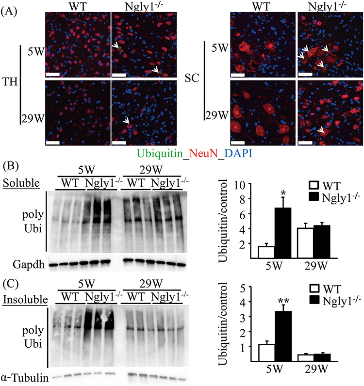
Figure 7.

Accumulation of ubiquitinated proteins in the central nervous systems of Ngly1−/− rats. (A) Ubiquitin-positive inclusions in neurons of Ngly1−/− rats. Immunohistochemistry of thalamus (TH) and spinal cord (SC) sections from Ngly1−/− and the WT rats at 5 and 29 weeks of age, stained with an anti-ubiquitin antibody and NeuN, a mature neuron marker. Arrows indicate ubiquitin-positive neurons. Nuclei were stained with DAPI. Scale bar 50 μm. (B, C) Accumulation of Triton-X-100-soluble (B) and insoluble (C) polyubiquitinated proteins in spinal cords of Ngly1−/− and WT rats. The total protein extracts were separated from the spinal cords of the rats and were separated into Triton-X-100-soluble and Triton-X-100-insoluble fractions and analyzed by immunoblotting using anti-polyubiquitinated antibodies (top) and anti-GAPDH or α-tubulin antibodies (bottom; loading control). Semi-quantitative analyses by densitometry were carried out (B, C). Values represent mean ± SEM (n = 3). Asterisks indicate *P < 0.05 and **P < 0.01.
