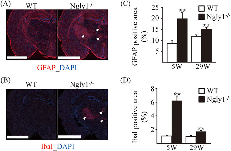
Figure 6.

Glial activation in thalamic VPL/VPM, VM and VL regions of Ngly1−/− rats. Immunohistochemistry for GFAP in thalamus of WT and Ngly1−/− rats at 5 and 29 weeks of age, showing an increase in area of reactive astrocytes. (A, B) Immunohistochemistry for GFAP (A) and IbaI (B) in the thalamus of WT and Ngly1−/− rats at 5 weeks of age. Scale bar 2.5 mm. Arrows indicate the regions with gliosis in Ngly1−/− rats. Nuclei were stained with DAPI. (C, D) Quantitative analyses show the area occupied by the GFAP (C) or IbaI (D) positive areas in thalamus of WT and Ngly1−/− rats. Values represent mean ± SEM (n = 6). Asterisks indicate **P < 0.01.
