
Figure 2.
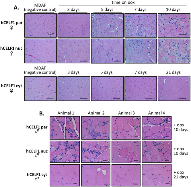
Parental and nuclear but not cytoplasmic mCherry-CELF1 transgenic lines develop severe histopathology. Adult hCELF1par, hCELF1nuc and hCELF1cyt bitransgenic mice were induced to express mCherry-CELF1 for the times indicated. (A) Panels show H&E staining of formalin-fixed, paraffin-embedded female quadriceps. MDAF alone control littermates on dox for 7 days show normal histology. Induced animals from the hCELF1par and hCELF1nuc lines have multiple histological defects such as myofiber variability, hyperpigmentation and myofiber necrosis. Animals expressing hCELF1cyt do not show evidence overt histopathology. Scale bar, 100 μm. (B) Panels for each line show H&E staining from four different male animals. hCELF1par and hCELF1nuc animals were induced for 10 days and hCELF1cyt animals were induced for 21 days. Examples of severe histological defects observed in animals expressing high levels hCELF1par and hCELF1nuc of mCherry-CELF1. Mice expressing high levels of hCELF1cyt show normal histology, with some areas of adipose deposition and myofiber variability but otherwise display no detectable histological defects 21 days post-induction. Scale bar, 25 μm.