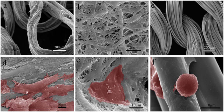
Figure 8.
Representative microscopic images of the collagen and PLA substrates and HUVECs attached at Day 3. (a) COL-K shares a similar structure with (c) PLA-K, but PLA-K has a smoother surface and larger surface area compared to COL-K due to the multifilament feature; (b) COL-KE, on the other hand, had a different morphology. A larger number of well-attached HUVECs grew on the collagen substrate (d), (e) compared to the PLA scaffold (f). Cells have been highlighted in red.

