
Fig. 1.
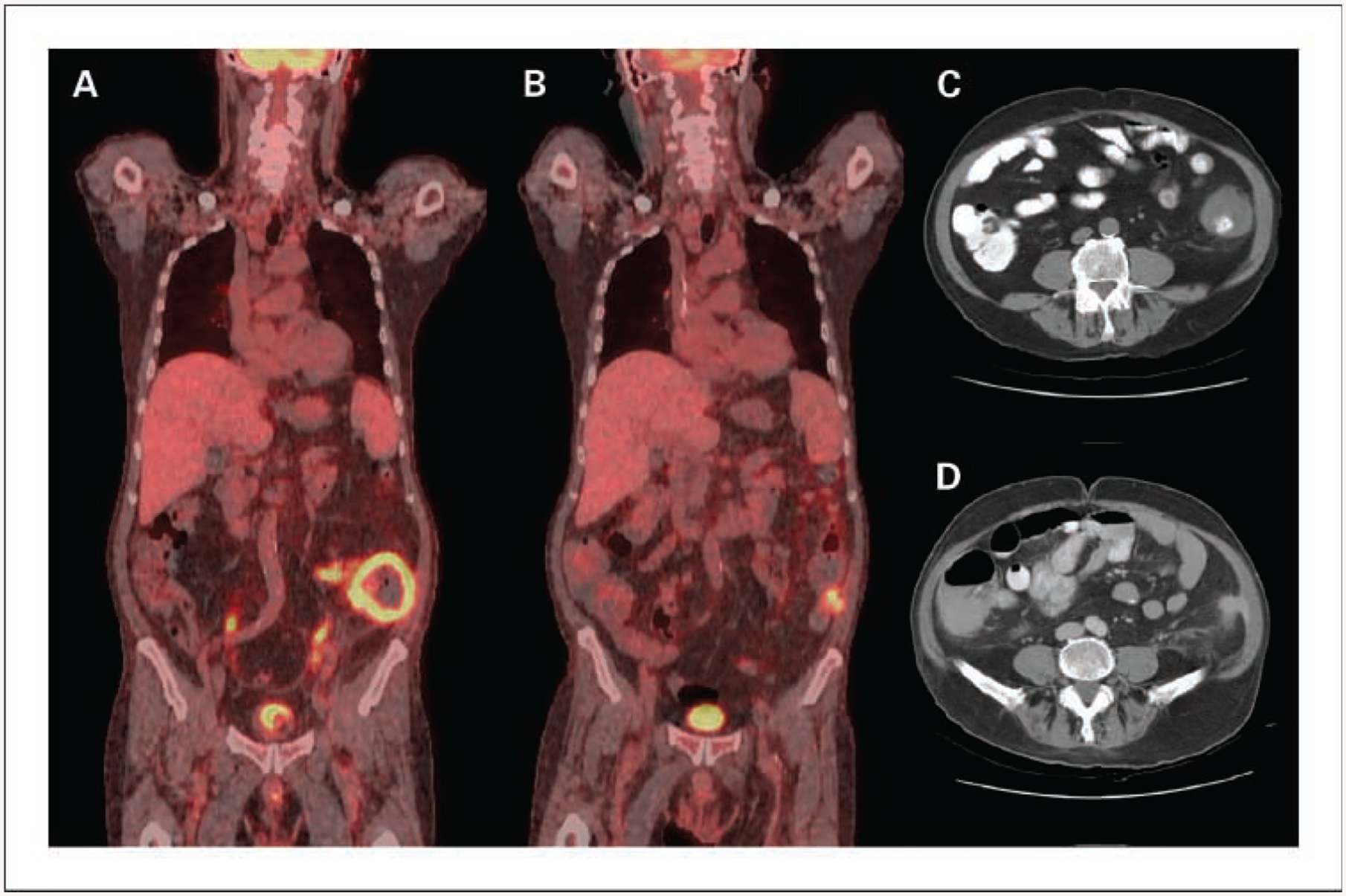
Radiographic imaging of the 72-year-old patient with extranodal non-Hodgkin’s lymphoma of gastrointestinal tract. A, sagittal 18-fluorodeoxyglucose-positron emission tomography image at diagnosis of EBV lymphoma showing circumferential metabolic activity within the wall of the descending colon. B, sagittal 18-fluorodeoxyglucose-positron emission tomography imaging after completion of chemotherapy showing small residual focus of metabolic activity. Computed tomographic images showing left lower quadrant mass involving descending colon at diagnosis (C) and imaging post-therapy shows minor residual abnormality (D).