
Fig. 3.
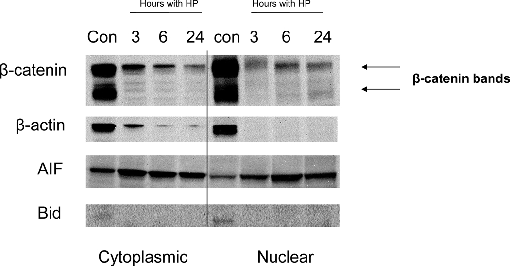
H. pylori-induced release of AIF. Western blots of AIF expression in cytosolic and nuclear fractions at 3, 6, 24 h after AGS treatment with H. pylori. Translocation of AIF from mitochondria to the cytoplasm and nucleus periphery was observed. Release of AIF into the cytoplasmic fraction peaked within 3 h, and translocation of AIF to the nucleus peaked at 6 h. The increase in cytoplasmic AIF occurred despite an apparent loss of β-actin. A band corresponding to full-length Bid was seen both in the cytoplasmic and nuclear fractions of controls, but not in cells treated with H. pylori. A time-dependent decrease in β-catenin protein expression was detected in cytoplasmic and nuclear extracts, as seen for β-actin, suggesting concentration-dependent protein cleavage with induction of apoptosis. H. pylori-induced apoptosis in AGS gastric cells involves cleavage of Bid. Immunoblot showing the presence of Bid in cells untreated cells. Cleaved Bid appeared only in the AGS cells treated with H. pylori. Cytoplasmic and nuclear proteins were run on SDS-PAGE and probed with anti-human Bid.