
Fig. 4.
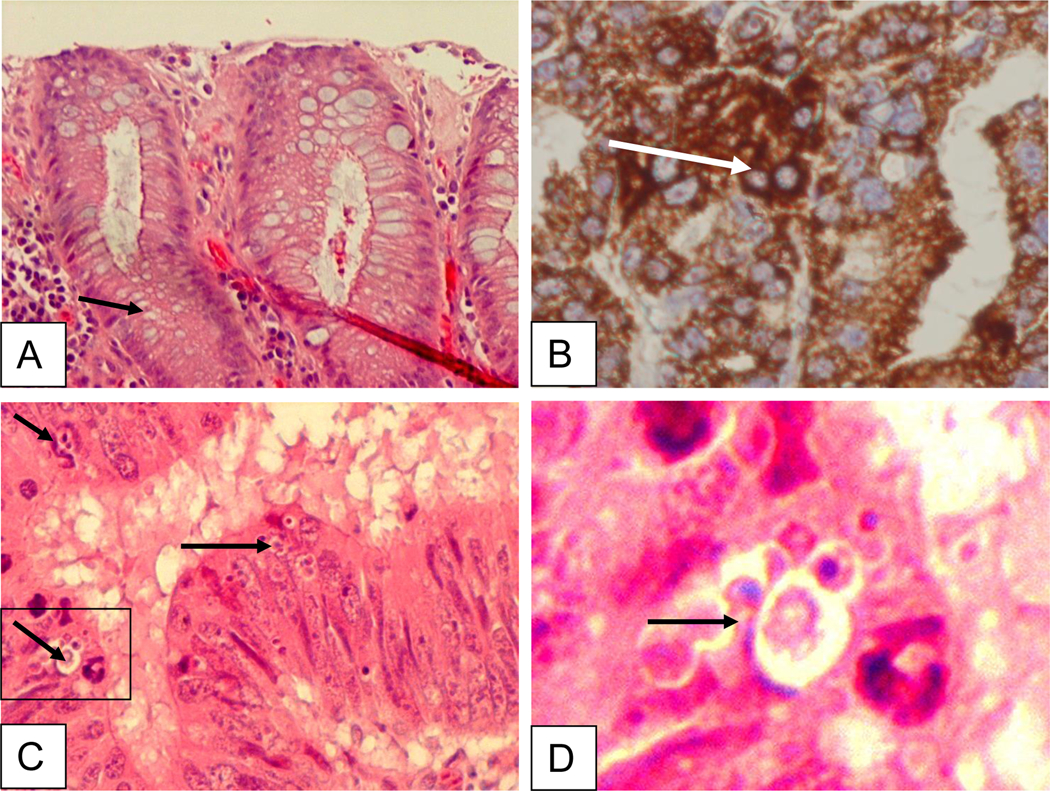
Immunohistochemical staining of gastric tissue infected with H. pylori. Bright field images of gastric tissue infected with H. pylori (B-D) vs. control (A). The tissue was stained with H & E stain, arrows show healthy (A), AIF cytoplasmic staining (B) or apoptotic cells (C, D). Cells were immunostained with primary AIF-specific antibody, (AIF goat polyclonal, clone D-20; sc-9416; Santa Cruz Biotechnology) and donkey anti-goat horseradish peroxidase conjugated secondary antibody for DAB staining. Adequate antibody penetration and cell architecture preservation is evidenced by cytoplasmic staining.