
Fig. 5.
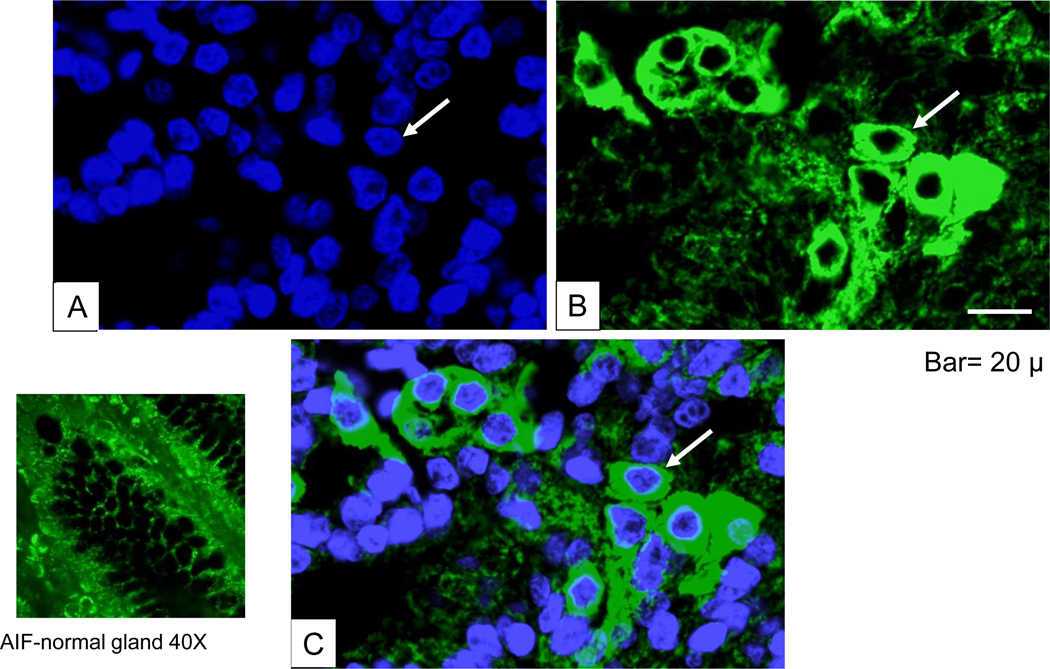
Immunofluorescent analysis of AIF. (A) DAPI-staining to visualize nucleus. (B) Immunohistochemical staining with rabbit polyclonal AIF primary antibody (sc-9417) followed by FITC-conjugated donkey anti-goat secondary antibody. (C) Merged image for viewing colocalization of AIF with DAPI-stained cytoplasmic and nuclear periphery staining. Bar =50 μm.