
Fig. 6.
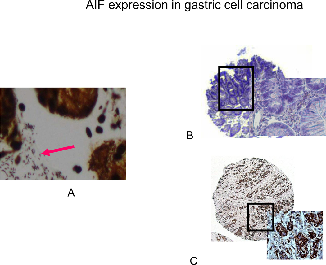
Silver (A) and Immunohistochemical staining of H. pylori and AIF in human tissue microarray (B, C). (A) Positive silver staining for H. pylori is evident in all of the glands in biopsy specimens from patients infected with H. pylori. (B) In uninfected patients, lack of brown color indicates absence of cytoplamic staining for AIF. (C) In patients infected with H. pylori, 28/44 (63%) of the cases showed strong cytoplasmic staining for AIF in the malignant epithelial cells.