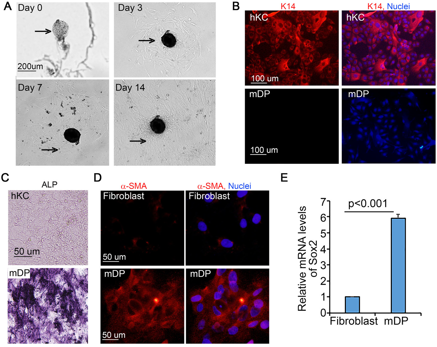
Figure 1. Passaged mouse dermal papilla (mDP) cells retained DP cell characteristics.

(A) Growth of mDP cells from DP condensates. Bright-field images were taken on day 1, 3, 7 and 14 after DP isolation. Arrows depict isolated DP condensates. (B) Immunostaining of human keratinocytes (hKC) and mDP cells with a primary antibody against cytokeratin 14 (K14) followed by detection with a Dylight 555-conjugated donkey anti-rabbit secondary antibody [orange]. Nuclei [blue, Hoechst 33258]. (C) Alkaline phosphatase staining of passaged mDP cells and hKC cells. (D) Immunostaining of passaged mDP cells and dermal fibroblasts for α-smooth muscle actin (a-SMA) [orange], Nuclei [Hoechst 33258, blue]. (E) Quantitative RT-PCR. Total RNA samples were isolated from cultured mDP and mouse dermal fibroblasts. Graph represents relative Sox2 mRNA levels of triplicate samples +/−SD. GAPDH was used for internal control.
