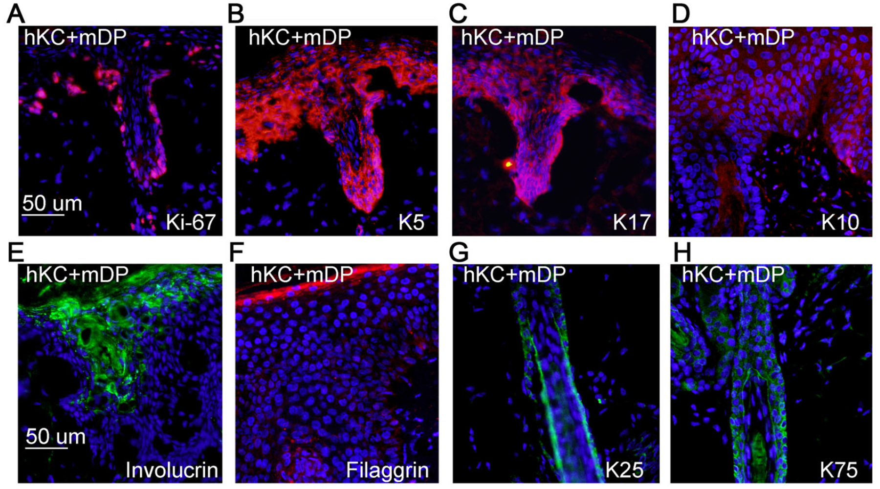
Figure 3. Regenerated hair follicles and epidermis were proliferative, and expressed relevant differentiation markers.

(A-E) Frozen tissue sections of 6-weeks old skin grafts regenerated with hKC and mDP cells were incubated with primary antibodies against Ki-67, K5, K17, K10, or filaggrin, and then detected with a Dylight 555-conjugated secondary antibody [orange], Nuclei [blue]. (F-G) Paraffin-embedded tissue sections of 6-weeks old skin grafts were incubated with primary antibodies against cytokeratin 25 (K25) and 75 (K75), and then detected with a Dylight 488-conjugated secondary antibody [green], Nuclei [blue].
