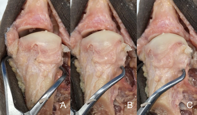
Figure 1.
Photograph of the cadaveric specimen demonstrating the first metatarsal bone dissected free from soft tissues from dorsal aspect. A clamp is used to apply pronation to the metatarsal bone. A, The metatarsal bone in an anatomic position, without rotation applied. B, The same metatarsal bone with 15° of pronation applied to it. C, The same cadaveric bone with 30° of pronation applied. Note the change in the silhouette of the lateral aspect of the metatarsal head as the metatarsal condyle starts to appear in the view. As the pronation increases, the lateral aspect of the metatarsal head changes from a straight angle corner to a rounded corner, appearance given by the metatarsal condyle.

