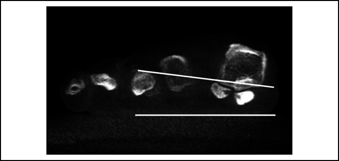
Figure 5.
Photograph demonstrating the weight-bearing CT scan of the same patient. There is no subluxation of the lateral sesamoid. Two lines have been drawn to delineate the floor and the metatarsosesamoid facets, which demonstrate the pronation of the metatarsal bone. Owing to the pronation, when looking on the anterioposterior radiographic image, a seudosubluxation of the sesamoids is seen, and we start to perceive a roundness of the lateral aspect of the metatarsal head.

