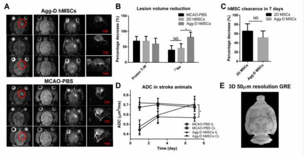
Figure 6.
In vivo MRI demonstrated stroke lesion recovery and cell clearance after transplantation of Agg-D hMSCs in MCAO animals. (A): Representative in vivo MRI image of T2-weighted, 2D GRE, 3D-23Na, and 2D diffusion weighted echo planner image (EPI) of Agg-D hMSCs and MCAO-PBS group at 1, 3, and 7 d after MCAO and injection. (B): Lesion volume reduction calculated from proton T2 weight and 23Na image. (C): Cell clearance of Agg-D and 2D hMSCs 7 days after transplantation. (D): Apparent diffusion coefficient (ADC) of Agg-D hMSCs and MCAO-PBS group. (E): Ex vivo, high-resolution (100-μm isotropic) 3D GRE images of a perfused MCAO rat that was injected with Agg-D hMSCs showing the retention of MPIO-labeled cells after one week. N=7 for 2D and Agg-D hMSC group, N=5 for MCAO-PBS group. NS, no significance; *, p<0.05.

