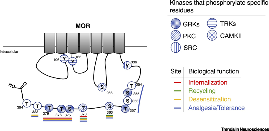
Figure 3: Biological relevance of µ-Opioid Receptor phosphorylation.

A schematic representation of the intracellular regions of rat µ-Opioid Receptor showing phosphorylatable residues and their biological relevance. Kinases that modify individual residues are indicated with filled hatches. The residue number is indicated next to each residue. Filled circles represent known phosphorylation sites and open circles represent putative phosphorylation residues. Residues that affect distinct biological activities are indicated with a colored line; in red are the residues described to play a role in MOR internalization; in green are the residues described to play a role in MOR recycling; in yellow are the residues described to play a role in MOR desensitization; and in blue are the residues described to play a role in opioid analgesia and tolerance. GRKs, G protein-coupled receptor kinases; RTKs, receptor tyrosine kinases; PKC, protein kinase C; CAMKII, Ca++/calmodulin kinase II; Src, Src kinase.
