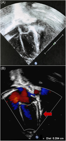
Figure 2.

A, Four chamber view of the heart taken during the first cardiological evaluation shows normal structure and dimensions of cardiac chambers, without pericardial effusion. B, Four chamber view of the heart four days after the previous one shows a 2 to 3 mm pericardial effusion in the lateral‐posterior pericardial space (arrow)
