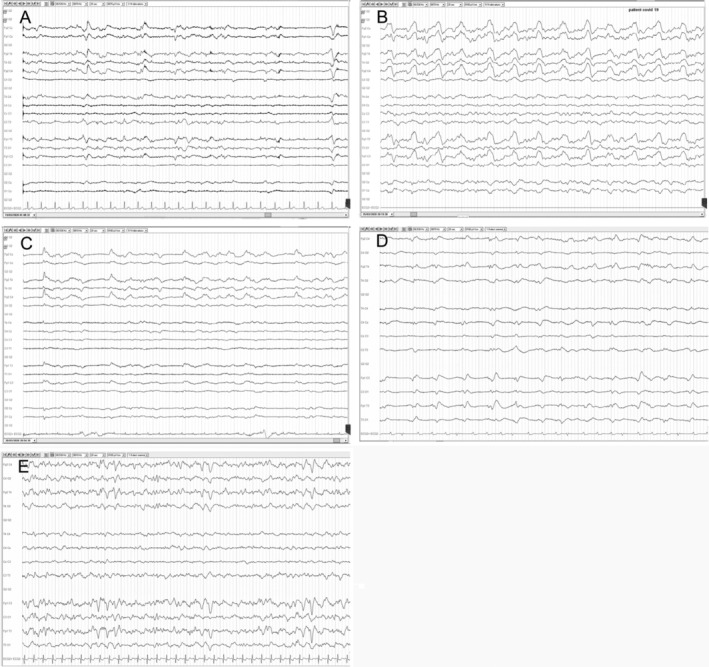
FIGURE: 1.

Electroencephalographic (EEG) recordings of Patients 1 through 5. The panels display EEG recordings of 20 seconds for each patient. In each panel, the EEG channels are shown with an electrocardiographic recording at the bottom. Nomenclature is in accordance with the International 10–20 montage. The EEGs reveal a pattern of slow waves with periodic discharges in the frontal regions. (A) Scalp EEG in a reference montage from Patient 1 reveals generalized rhythmic delta activity (GRDA) with intermittent biphasic delta waves in bilateral frontal regions that are symmetric and monomorphic with low‐voltage rhythmic background activity. (B) Scalp EEG in a reference montage from Patient 2 reveals GRDA with frequent high‐amplitude biphasic delta waves in bilateral frontal regions that are symmetric and polymorphic with low‐voltage rhythmic theta background activity. (C) Scalp EEG in a reference montage from Patient 3 reveals lateralized periodic discharges of high‐amplitude monomorphic delta activity with right frontal region predominance and low‐voltage rhythmic theta background activity. (D) Scalp EEG in a reference montage from Patient 4 reveals GRDA with intermittent low‐amplitude slow biphasic delta waves in bilateral frontal regions that are slightly asymmetric and monomorphic with low‐voltage continuous background activity. (E) Scalp EEG in a reference montage from Patient 5 reveals GRDA with intermittent high‐amplitude biphasic delta waves in bilateral frontal regions that are symmetric and monomorphic with intermittent low‐voltage rhythmic theta background activity.
