Figure 4.

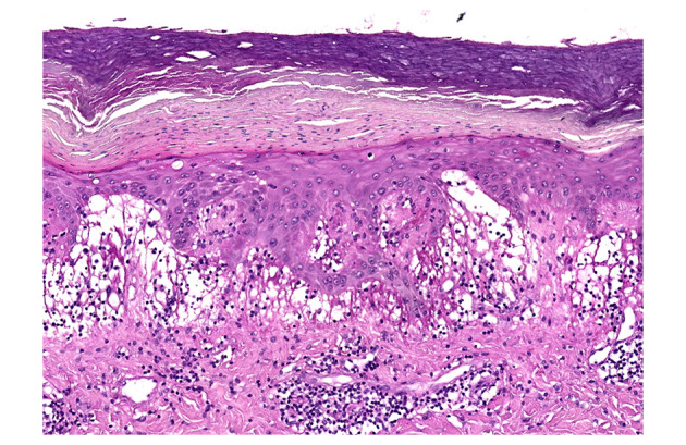
Histology. Dilated vessels, massive oedema, marked infiltrate lymphocytes in the papillary dermis. Dense infiltrate of lymphocytes at the dermoepidermal junction. Some necrotic keratinocytes in the epidermis. H&E 10×.

Histology. Dilated vessels, massive oedema, marked infiltrate lymphocytes in the papillary dermis. Dense infiltrate of lymphocytes at the dermoepidermal junction. Some necrotic keratinocytes in the epidermis. H&E 10×.