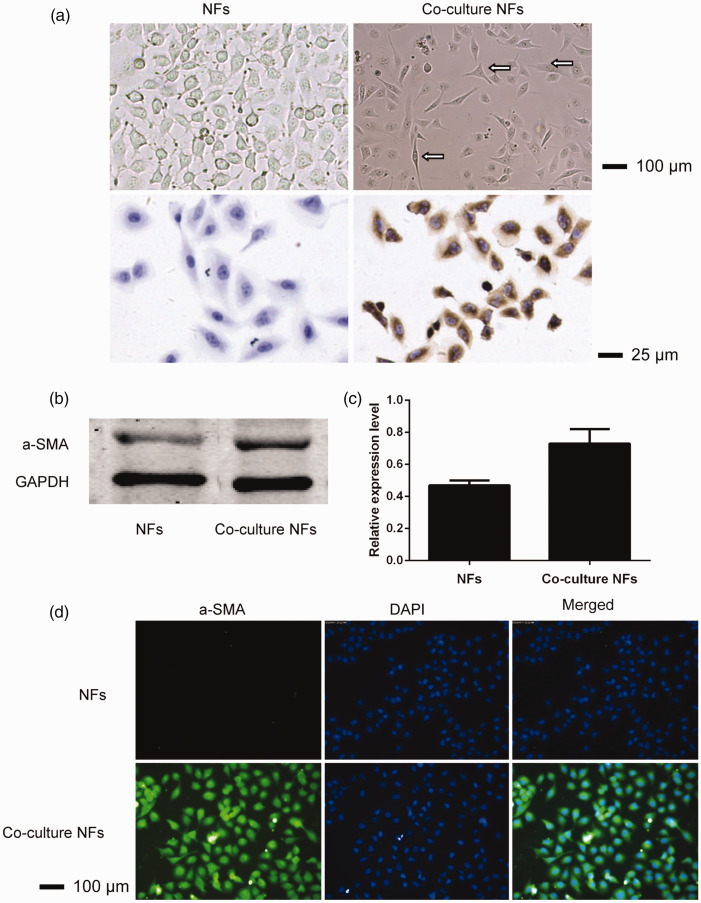
Figure 1.
Morphological analysis (first line, ×100) and chemical staining (second line, ×400) of NFs and co-culture with NFs from colon cancer (a). NFs showed fusiform or star-shaped morphology of the same size, while after co-culture, the morphology of the NFs was fusiform or a polygon with different sizes. This could be observed in the binuclear and polynuclear cells, while α-SMA expression was negative in NFs and positive in co-culture NFs. α-SMA protein expression in NFs and co-cultured NFs from colon cancer was determined using SDS-PAGE and an ODYSSEY fluorescence imaging system (b); calculations were performed using ImageJ image analysis software (c); and the cellular immunofluorescence results are presented (d).
NF, normal fibroblasts; α-SMA, α-smooth muscle actin; SDS-PAGE, sodium dodecyl (lauryl) sulfate-polyacrylamide gel electrophoresis.

