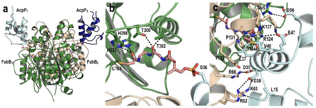
Figure 1: Crystal structure of E. coli AcpP-FabB complex.

(a) Overall AcpP-FabB complex structure, with the FabB monomers shown in dark green and light tan, the AcpP monomers shown in dark blue and light cyan, and the crosslinker in pink. AcpP helices are labeled I - IV. (b) FabB active site interactions with the crosslinker. (c) AcpP-FabB interface interactions.
