
Figure 7.
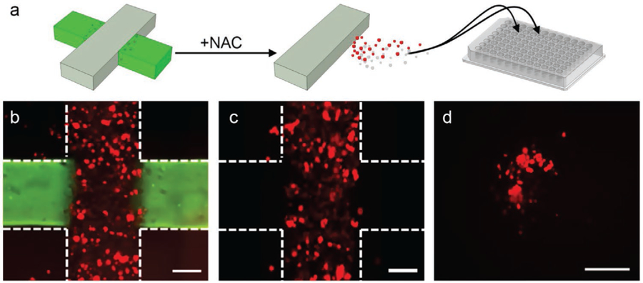
Dissolving of PEGSSDA hydrogel domains. a) Schematic representation of selective recovery of infiltrated cells (red) from a cross structure (left). Non-infiltrating cells in grey region not shown for clarity. NAC is used to dissolve the gel in the cell-free domain (green), releasing only invaded cells into circulation (center) where they can then be recovered and loaded onto a 96-well plate (right) for analysis. Overlaid 2D images of the multi-domain construct on day 10 before (b) and after (c) dissolving the gel with NAC. The vertical cell-laden domain encapsulates mCherry cells (red) and the horizontal domain is stained with Alexafluor 488 for structure visualization. Construct borders are roughly indicated by white dashed lines and scale bars are 300 µm. Following NAC (c), the green fluorescent region (hydrogel with PEGSSDA crosslinker) is selectively removed along with any infiltrated cells it contained. d) 2D image of cells recovered from the construct in a 96-well plate and incubated for 4 h. Scale bar is 300 µm.