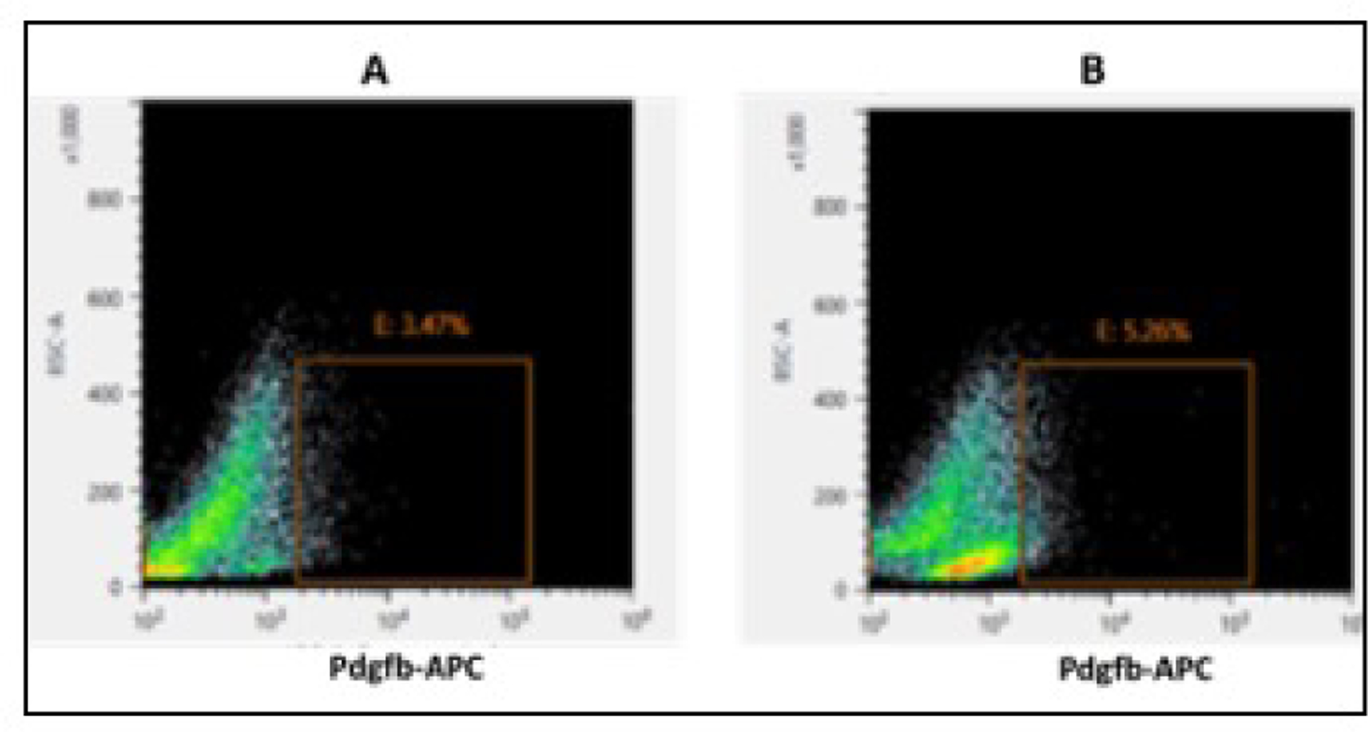
Figure 2.

FACS purification of retinal pericytes. The representative Dot blots display the intensity of channel of analyzed retinal dissociated cells for Pdgfrb-APC staining from (A) non-diabetic mice retina and (B) diabetic mice retina (12 weeks old). The sorted, Pdgfrb-APC positive cell population for pericytes is specified by the inset.
