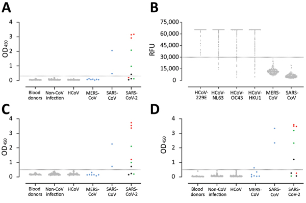
Figure 3.

Validation of use of S1 (A, B), RBD (C), and N protein (D) ELISAs for detection of SARS-CoV-2–specific antibodies infections. Gray dots indicate specificity cohorts A–C, including healthy blood donors (n = 45), non-CoV respiratory infections (n = 76), and HCoV infections (n = 75); blue dots indicate non-SARS-CoV-2 zoonotic coronavirus infections (i.e., MERS-CoV [n = 7] and SARS-CoV [n = 2]); red dots indicate patients with severe COVID-19; and green and black dots indicate patients with mild COVID-19. Dotted horizontal lines indicate ELISA cutoff values. CoV, coronavirus; COVID-19, coronavirus disease 2019; HCoV, human coronavirus; MERS-CoV, Middle East respiratory syndrome coronavirus; N, nucleocapsid; OD, optical density; RBD, receptor-binding domain; RFU, relative fluorescence unit; S, spike; SARS-CoV, severe acute respiratory syndrome coronavirus; SARS-CoV-2; severe acute respiratory syndrome coronavirus 2.
