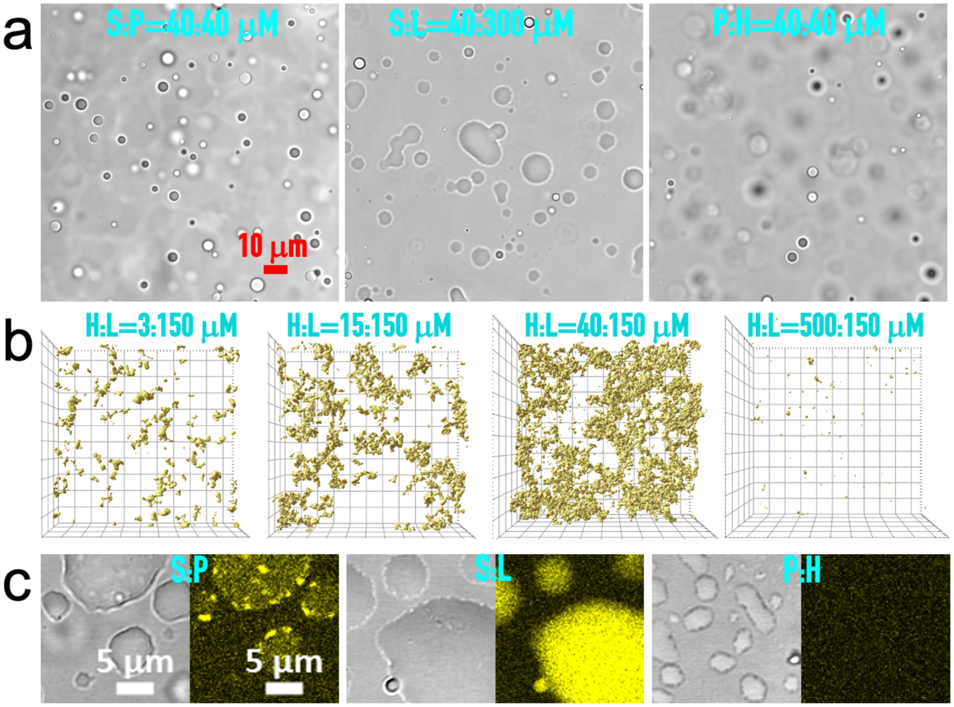
Figure 1. Phases of binary mixtures.

(a) Brightfield images of droplets formed by S:P, S:L, and H:P binaries, at concentrations indicated. The image of S:P droplets has appeared previously50. (b) Z stacks of confocal images of H:L precipitates taken at approximately 20 min after mixing H at four concentrations with L at 150 μM, visualized by the fluorescence of thioflavin T (ThT) in a top view. The field of view is 105 μm × 105 μm and the height of the Z stacks range from 30 to 50 μm. (c) Brightfield and fluorescence images of S:P, S:L, and H:P droplets, showing moderate, strong, and no ThT binding (yellow), respectively. Concentrations were S:P = 40:40 μM, S:L = 20:300 μM, and P:H = 10:40 μM. Images were taken at 10 to 20 min after mixing; by then droplets had fallen, fused, and spread over a coverslip.
