
Figure 1.
Obesity Exacerbates COVID-19: Potential Mechanisms
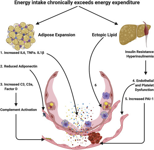
Obesity is a disorder of energy balance that ensues when energy intake exceeds expenditure. Adipose tissue expansion occurs to safely store excess energy safely in triglyceride-rich lipid droplets. This process is associated with adipose tissue inflammation and elaboration of pro-inflammatory cytokines, increased components of the complement system, and altered adipose tissue hormones.
(1) Increased inflammatory cytokines are secreted into the systemic circulation and can act on the alveolar capillary unit to potentiate the inflammatory response to SARS-CoV-2 infection.
(2) Adipose tissue expansion is associated with a reduction in adiponectin secretion from the adipose tissue that is at least partly driven by systemic insulin resistance. Mouse studies suggest that adiponectin is abundant in the pulmonary endothelium in the healthy lung and that adiponectin deficiency results in pulmonary vascular inflammation and predisposes to experimental lung injury.
(3) Increases in circulating complement components elaborated from adipose tissue occur in expanded adipose tissue and in association with insulin resistance and could predispose to complement activation and subsequent thrombotic microangiopathy. When the capacity for adipose tissue to expand is exceeded, lipid is deposited in other organs. Lipid deposition in the skeletal muscle and liver likely plays a causal role in the development of insulin resistance and hyperinsulinemia.
(4) Systemic insulin resistance is associated with endothelial and platelet dysfunction that may predispose to thrombosis and contribute to lung injury via vascular inflammation and enhanced endothelial permeability.
(5) Insulin resistance is robustly associated with increased plasminogen activator inhibitor-1 (PAI-1), which impairs fibrinolysis and may contribute to the risk of thrombosis in COVID-19.
(6) Finally, ectopic lipid may actually be directly deposited in type 2 pneumocytes pre-disposing to lung injury in SARS-CoV-2 infection.