Figure 1.

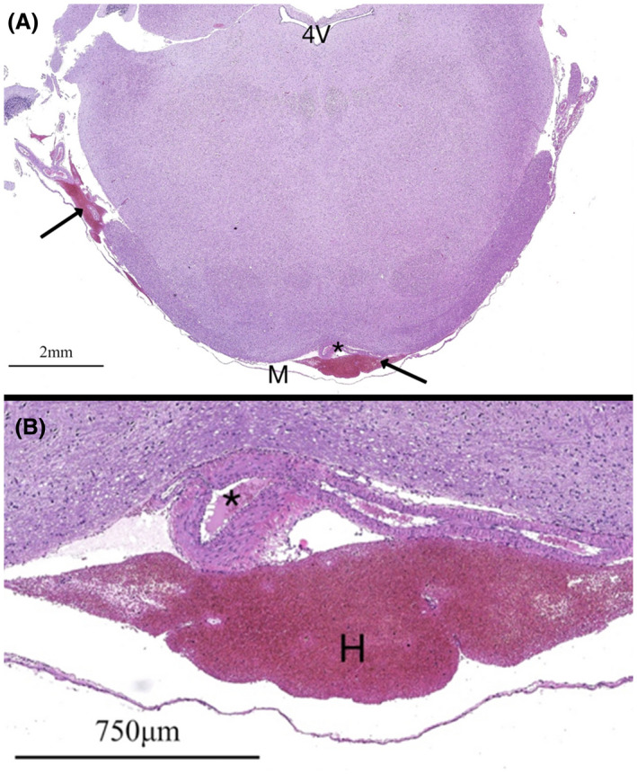
H&E‐stained specimens from a NZW rabbit (animal 13) at the level of the brainstem and fourth ventricle (4V) at 10× (A) and 200× (B) magnification. A decompressed normal basilar artery is noted (*), the lumen of which contains erythrocytes from poor clearance during perfusion. Subleptomeningeal hemorrhage (H, arrows) is noted from complications during fixation
