Figure 2.

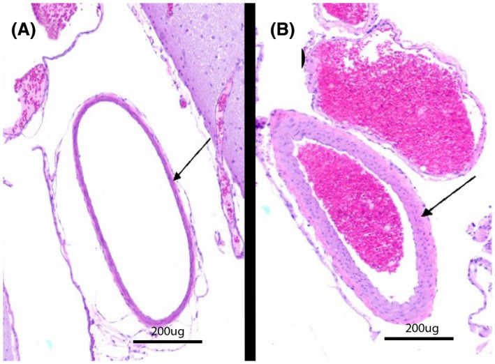
H&E‐stained specimens of left ICAs from two different ApoE‐KO rabbits with thin but normal (A, animal 6) and hypertrophied (B, animal 10) arterial walls, the latter of which is consistent with mild atherosclerotic changes

H&E‐stained specimens of left ICAs from two different ApoE‐KO rabbits with thin but normal (A, animal 6) and hypertrophied (B, animal 10) arterial walls, the latter of which is consistent with mild atherosclerotic changes