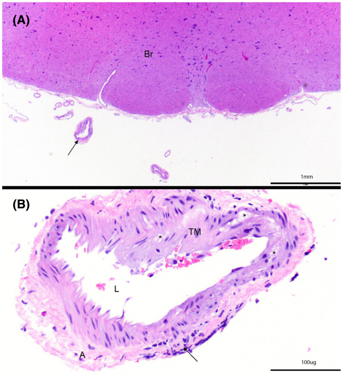
Figure 3.

H&E‐stained specimen from an ApoE‐KO rabbit (animal 8) at (A) 20× and (B) 200× at the level of the brainstem (Br) demonstrate expansion of the tunica media (TM) by vacuolated cells (*). Black arrow indicates inflammatory cell infiltration. Adventitia (A) and lumen (L) are also indicated. Note, image B is rotated clockwise compared to image A. Arterial findings are consistent with moderate atherosclerosis
