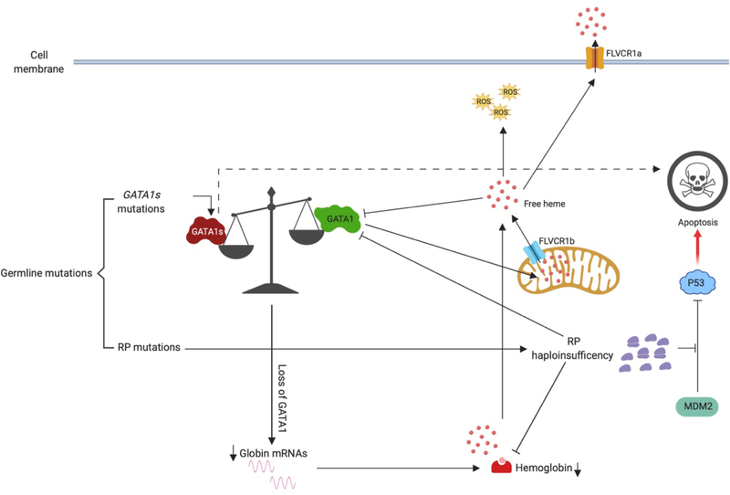
Figure 2. Mechanisms that lead to Diamond-Blackfan anemia.
The majority of germline mutations found in DBA patients reside in ribosome protein (RP) coding genes. The resulting RP haploinsufficiency leads to inefficient translation of a number of erythroid genes, such as globin and GATA1, resulting in impaired erythropoiesis. Moreover, free ribosomal subunits, such as RPL5 and RPL23, block MDM2-mediated P53 ubiquitination and degradation, leading an increase of P53-dependent apoptosis of the erythroid progenitors. In other patients, GATA1 gene mutations result in loss of the full-length protein but allow for expression of the shorter isoform (GATA1s mutations), which also impairs erythropoiesis. Finally, an altered globin-heme balance has been shown to lead to the accumulation of free heme in cytoplasm, which downregulates GATA1 at both the mRNA and protein level. FLVCR1a and FLVCR1b are heme transporters on cell membrane and mitochondrial membrane respectively.

